
News
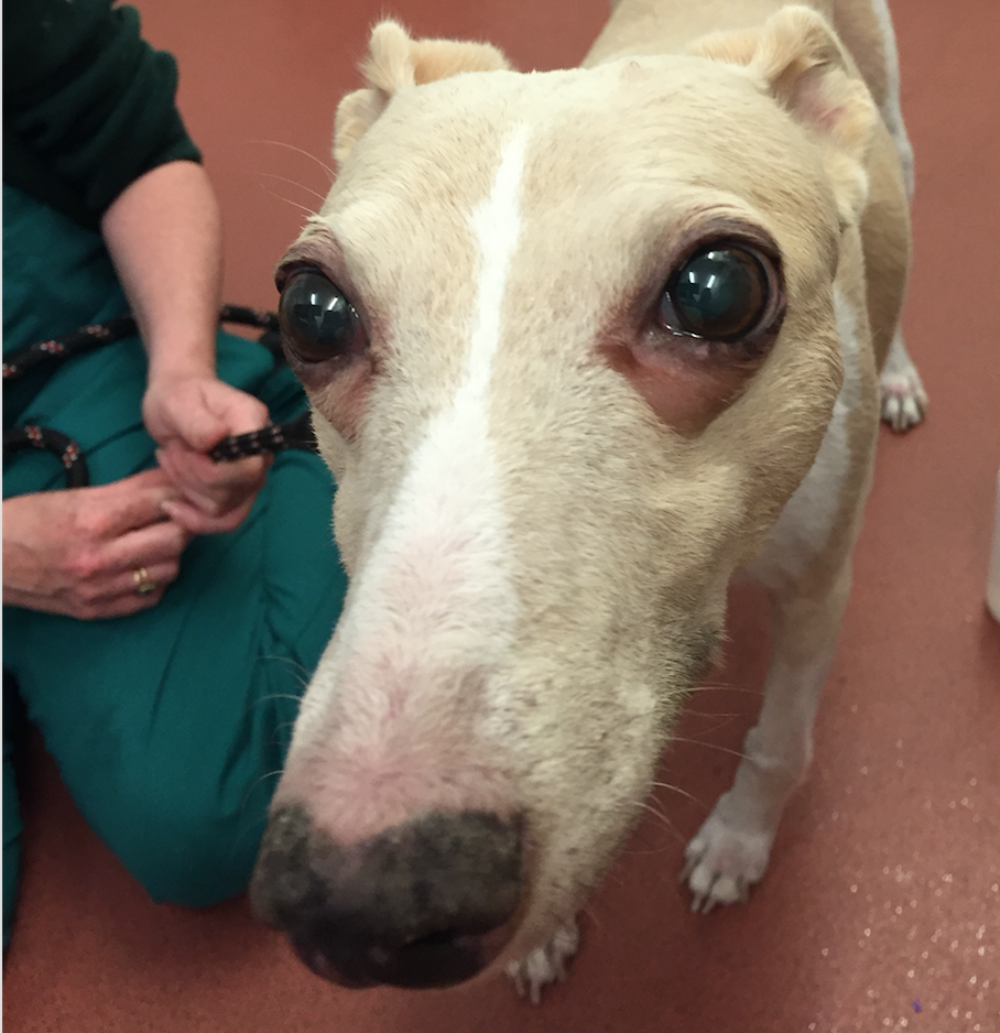
Pet of the Month – June 2016 – Polly
by admin on June 1st, 2016
Category: Pet of the Month, Tags:
What is Cushing’s disease and how is steroid produced by the body?
Hyperadrenocorticism or ‘Cushing’s disease’ is caused by excessive production or administration of steroid. It can be classified as pituitary dependent, adrenal dependent, or iatrogenic.
A brief overview of how steroid is produced in the body will help with the understanding of this disease. The pituitary gland is a small gland at the base of the brain that secretes a hormone called ACTH. ACTH causes the adrenal glands to secrete steroid hormone. There are two adrenal glands in animals which are located in the abdomen close to the left and right kidneys. The main steroids produced in response to ACTH secreted by the pituitary gland are glucocorticoids and these have widespread effects on the management of proteins and sugars by the body.
What is the cause of Cushing’s disease?
Approximately 85% of dogs with Cushing’s disease have a very small, typically non-cancerous, growth of the pituitary gland that drives the excessive steroid production (via excessive ACTH production). These dogs have what is called ‘pituitary dependent hyperadrenocorticism’ (abbreviated to ‘PDH’). Approximately 75% of dogs with PDH weigh less than 20kg.
Approximately 15% of dogs have a tumour on one of their adrenal glands that drives the excess steroid production. This is known as ‘adrenal dependent hyperadrenocorticism’ (abbreviated to ‘ADH’). Cancerous and non-cancerous growths occur with equal frequency. Approximately 50% of dogs with ADH weigh greater than 20kg.
What are the signs of Cushing’s disease?
The most common presenting signs in dogs with Cushing’s disease are:
- increased drinking
- increased urination
- increased appetite
- panting
- enlargement of the abdomen
- hair loss
Dogs with Cushing’s disease are usually 6 years of age or older and many different breeds have been reported to be affected including Poodles, various Terrier breeds, Daschunds, Beagles and Labrador Retrievers.
How is a diagnosis of Cushing’s disease made?
The diagnosis of Cushing’s disease can sometimes be difficult, even with blood tests. Blood test results will be interpreted by your vet in light of the history and findings on examination. A blood test taken after the administration of a hormone or drug is necessary to help make the diagnosis. These tests are called the ‘ACTH stimulation test’ and the ‘low dose dexamethasone suppression test’. Normally only one test is necessary; however, occasionally both tests need to be performed. Urine tests are often performed as well.
Once a diagnosis of Cushing’s disease is made, it is useful to know whether the patient has pituitary dependent Cushing’s disease or adrenal dependent Cushing’s disease. The differentiation can be made with further blood testing or with an ultrasound scan of the abdomen. An ultrasound scan of the abdomen is sometimes required to help in achieving a diagnosis of Cushing’s disease.
How are dogs with Cushing’s disease treated?
Dogs with confirmed pituitary dependent Cushing’s disease are managed with an oral drug called trilostane. Trilostane stops the production of steroid and leads to an improvement in clinical signs in most dogs. Trilostane is absorbed better if given with food. Blood testing is necessary after starting treatment to monitor the effect of the drug. Any blood test result will always be interpreted in combination with the clinical signs. The majority of dogs will show a good response to treatment but will require lifelong therapy. Occasionally the small growth on the pituitary gland can grow and cause neurological signs (fits, behaviour change). Scans of the brain are therefore sometimes performed.
Surgical removal of the adrenal gland mass is the treatment of choice for patients with adrenal dependent Cushing’s disease. The outlook depends upon the degree of invasion of the growth into local blood vessels and analysis of the tumour under a microscope. Trilostane can be trialled in those patients for whom surgery is not an option.
‘Iatrogenic’ Cushing’s disease can be caused by the administration of steroid by mouth or topically. It tends to occur in patients on high dose, long term oral steroid (prednisolone) therapy. Treatment involves gradually stopping steroid therapy.
Management of Atopic Dermatitis in Dogs and Cats
by admin on June 1st, 2016
Category: News, Tags: Tags: atopic dermatits, cats, dogs, pets
Last month we explained what atopic dermatitis is and how it is diagnosed. In this article we consider how atopic dermatitis can be managed in affected dogs and cats.
Can I cure atopic dermatitis?
In one word “No”, but you can manage the condition successfully. Atopic dermatitis in dogs and cats can be compared to asthma in people. Asthma can’t be cured but it can be managed; and just like asthma the management of atopic dermatitis is life-long. It is therefore important to put in place measures that are going to have the least side-effects for your pet in the long term that will provide him/her a good quality of life and that are the most cost effective and affordable for you.
How can I manage the condition?
You need to take a multi-step approach to managing atopic dermatitis. These steps include:
- Treatment and prevention of infections
- Medications to stop the itch
- Treatment that modulates the immune system
- Nutritional supplements
- Topical treatments
- Unfortunately some treatments work some of the time but not all the time and so you may need to switch treatments periodically. Some treatments may result in undesirable side effects and so may need modification.
Management options:
1) Treatment and prevention of infections:
Any bacterial (mostly staphylococcal) and/or yeast (Malassezia) infections should be treated at the outset. Antibiotics for bacterial infections need to be administered for 7 – 14 days beyond clinical cure and the duration of treatment is generally determined by the depth of the pyoderma.
For yeast infections a shampoo containing either 2% chlorhexidine/2% miconazole or one containing 3% chlorhexidine have shown good efficacy and will suffice; however, if the infection is severe your vet may opt to prescribe antifungal tablets. Because there is a tendency for infections to recur it is best to bath your pet once or twice a week on a regular basis, which will keep the microbial load on the skin low and thus help reduce the frequency of infections.
2) Stopping the itch:
Glucocorticoids (steroids) will stop the itch in most cases, but need to be used with caution, especially if used for a long time. In the short term they can increase water intake, increase urination and appetite. Some owners and pets find these side effects distressing. In the long term they can affect almost any organ in the body, therefore, even when well tolerated, they should be used with caution and your pet should be monitored regularly. Topical steroid containing sprays and ointments may be useful for targeted areas but with long term use thinning of the skin, infections and systemic side effects can occur.
Cyclosporin is used to damp-down (modulate) an over-reacting immune system in an atopic pet. One starts with a high dose and then tapers it down to alternate day, or better still twice a week, treatment. The most common side effects with ciclosporin are vomiting and diarrhoea. In some dogs, gingival hyperplasia and papillomas may also occur.
Oclacitinib is another immunomodulating drug which specifically targets the pathway that results in an itch. It is effective and can stop itching rapidly. This is the newest drug on the market and the reported side effects are vomiting and diarrhoea in a small number of cases. It can be used both short term and long term; however, given that this drug has now been on the open market for only a few months (at the time of writing this) it should be used with caution as side effects with prolonged use are not known.
Antihistamines; the response to this group is variable and they are often used in combination with steroids in order to reduce the steroid dose.
3) Immune Modulation:
Allergen specific immunotherapy is the most specific treatment for Atopic Dermatitis. It involves either an injection, or an oral dose of the allergens your pet is allergic to, to modulate the immune system. It has responses ranging from roughly 33% where there is complete cessation of itch, to 33% improving but requiring additional treatments, to 33% where there is no response. If your pet responds well to immunotherapy, it is likely to have the least adverse side-effects and to be the most cost effective treatment in the long term.
4) Nutritional support:
Omega -3 and Omega -6 essential fatty acids help the skin function by altering the lipid barrier and by reducing inflammation. On their own they are unlikely to benefit your pet but can be helpful when combined with other treatments. Diets that are high in essential fatty acids (EFAs) also help.
5) Topical treatments:
Moisturisers containing ceramides help maintain the skin barrier, which in turn reduces the penetration of allergens and therefore your pet’s reaction to them. Bathing with an oatmeal based shampoo can also help relieve itching and re-hydrate the skin.
Atopic dermatitis is a lifelong condition that requires life-long management; and often more than one type of treatment will be required. So, when your vet chooses a treatment, it should be efficacious, suitable for your pet with the least side effects, easy for you to administer and affordable. Most of all it/they should improve your pet’s (and your) quality of life.
Pet of the Month – May 2016 – O’Riley!
by admin on May 2nd, 2016
Category: Pet of the Month, Tags:

With O’Riley already suffering from Inflammatory Bowel Disease (IBD) his owners were used to him having the occasional flare ups of IBD causing him to not be his normal bouncy self. But when they noticed he was drinking more, urinating more and having more frequent bouts of vomiting as well they brought him in to see if anything else could be contributing to this.
Blood tests showed O’Riley to have higher than normal levels of urea and creatinine, substances which the kidneys excrete, indicating O’Riley’s kidney function was declining. A urine sample was taken to help confirm the diagnosis and also to check if he had excess protein in his urine (‘proteinuria’) as animals with kidney disease who develop this will benefit from additional medications. A clinically silent urinary tract infection was detected, a common complaint for animals with kidney disease, which obscured the protein results. This was addressed with a course of antibiotics before repeating the urine test. O’Riley was found to have proteinuria and started on a medication called an ACE inhibitor. An ultrasound scan of his kidneys was also performed to check for any structural abnormalities and his left kidney was found to be small, a common finding in Chronic Kidney Disease (CKD). A renal diet which restricts the amounts of protein and phosphorus, which the kidneys struggle to excrete in kidney disease, was tried but due to O’Riley’s concurrent IBD this treatment had to be abandoned. His phosphate levels were found to be too high and so an additional medication known as a phosphate binder was started. O’Riley also had regular checks of his blood pressure, as hypertension (high blood pressure) is another common result of CKD. When O’Riley’s blood pressure was found to be elevated he was also started on Amlodipine to help reduce this. Blood tests to check on his kidneys a month later showed improvements with lower levels of urea, creatinine and phosphate.
We are delighted to report that apart from an isolated occasion when his kidney disease made him feel unwell and which a brief stay in our hospital on intravenous fluids helped rectify, six months on O’Riley continues to do well.

Atopic Dermatitis
by admin on May 2nd, 2016
Category: News, Tags: Tags: atopic dermatits, dog, skin, sore
What is atopic dermatitis?
Many of you will have been told by your vet that your pet has atopic dermatitis, but what is it? Atopic dermatitis is the visible sign of a hypersensitivity (allergic) reaction; specifically a reaction to something (called an allergen) in your pet’s environment, either indoors or outdoors. It may be a reaction to pollens, or moulds, or to mites, such as house-dust mites.To complicate the issue further, some pets can have an allergic reaction to some foods, which is ‘food induced atopic dermatitis’.
Hypersensitivity is when the immune system goes into an overdrive. Normally when a pet is exposed to an allergen, its immune system produces antibodies to the allergen and once it has performed its protective role it will automatically revert to normal; however, if your pet is predisposed to atopic dermatitis its immune system continues to produce antibodies, which may then result in a hypersensitivity.
What are the main signs of atopic dermatitis in dogs and cats?
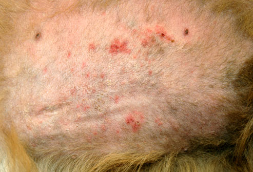
The hallmark of atopic dermatitis is an itch, usually without any visible signs other than a reddening of the skin. Often when I ask an owner where their pet animal is itching they say “Oh, Fido does not scratch”, but dogs exhibit itch by licking, rubbing, or chewing at the itch as well as by scratching at it.
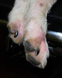
The itching usually starts between the ages of 6 months and 3 years. Initially it may be seasonal – that is it occurs only at certain times of the year – but over the years that can change. Spring and summer time itching is usually associated with pollen allergies (e.g. tree, grass and weed pollens) whereas winter or all year round itching is associated with indoor allergens such as house dust and storage mites or mould allergens. Food associated atopic dermatitis is likely to be non-seasonal, unless your pet is only being fed the food intermittently (e.g. on holiday). The itchy areas of the skin are usually the face, ears, feet and underside.
Often the condition is complicated with secondary infections (bacterial and/or yeast) and sometimes, just to complicate the situation further; some patients have been known to develop an allergy to the infection as well. Infections increase the level of itching and so just treating these will often decrease or stop the itch entirely. Once infections occur a rash may appear on the belly, neck and feet and your pet will lick, chew or scratch these areas. Infected skin is malodorous (has a bad smell) and, as the infection progresses the skin becomes thickened, blackened and crusty. If your pet then licks or scratches these areas aggressively, the skin can become broken or ulcerated, and also bleed.
Since similar lesions can also be seen with parasitic infestations (e.g. mange) these must be ruled out, or treated, at the same time as the allergy.
Recurrent ear infections are also associated with food or environment associated atopic disease. Usually the ear flap and the ear canals appear red in the early stage, which is then often followed by head shaking, discharge and swelling.
What is my pet allergic to?
To find out what your pet is allergic to, your vet can perform an allergy test, which will either involve sending a blood sample to a laboratory, or performing an intradermal skin test. Intradermal skin tests are when very small amounts of allergens are injected under the skin to identify which ones that particular animal is allergic to by provoking a small, controlled allergic response. Both forms of testing have their advantages and disadvantages. The results of the test should be interpreted bearing in mind the seasonality of the condition and your pet’s environment.
Allergy testing identifies the allergens to which your pet might be allergic to, but the main reasons for testing should be to find out what your pet is allergic to, so as to be able to improve quality of his/her life. Your vet can use the information for therapeutic purposes and to look at means of reducing the allergen load in the environment. The lower the allergen load, the lesser the itch and the easier it will be to keep your pet comfortable. One can compare atopic dermatitis in dogs to hay fever in people. If you have hay fever and you stand in a field full of flowers whose pollen you are allergic to, you will suffer severely, as opposed to if you stayed indoors when the pollen count is high. The same goes for dogs, by reducing the allergen load you will be helping to relieve your pet’s symptoms.
Can I cure atopic dermatitis?
In one word “No”, but you can manage the condition successfully. Atopic dermatitis in dogs and cats can be compared to asthma in people. Asthma can’t be cured but it can be managed; and just like asthma the management of atopic dermatitis is life-long. It is therefore important to put in place measures that are going to have the least side-effects for your pet in the long term that will provide him/her a good quality of life and that are the most cost effective and affordable for you.
How can I manage the condition?
You need to take a multi-step approach to managing atopic dermatitis. These steps include:
- Treatment and prevention of infections
- Medications to stop the itch
- Treatment that modulates the immune system
- Nutritional supplements
- Topical treatments
Unfortunately some treatments work some of the time but not all the time and so you may need to switch treatments periodically. Some treatments may result in undesirable side effects and so may need modification.
Management options:
1) Treatment and prevention of infections:
Any bacterial (mostly staphylococcal) and/or yeast (Malassezia) infections should be treated at the outset. Antibiotics for bacterial infections need to be administered for 7 – 14 days beyond clinical cure and the duration of treatment is generally determined by the depth of the pyoderma.
For yeast infections a shampoo containing either 2% chlorhexidine/2% miconazole or one containing 3% chlorhexidine have shown good efficacy and will suffice; however, if the infection is severe your vet may opt to prescribe antifungal tablets. Because there is a tendency for infections to recur it is best to bath your pet once or twice a week on a regular basis, which will keep the microbial load on the skin low and thus help reduce the frequency of infections.
2) Stopping the itch:
Glucocorticoids (steroids) will stop the itch in most cases, but need to be used with caution, especially if used for a long time. In the short term they can increase water intake, increase urination and appetite. Some owners and pets find these side effects distressing. In the long term they can affect almost any organ in the body, therefore, even when well tolerated, they should be used with caution and your pet should be monitored regularly. Topical steroid containing sprays and ointments may be useful for targeted areas but with long term use thinning of the skin, infections and systemic side effects can occur.
Cyclosporin is used to damp-down (modulate) an over-reacting immune system in an atopic pet. One starts with a high dose and then tapers it down to alternate day, or better still twice a week, treatment. The most common side effects with ciclosporin are vomiting and diarrhoea. In some dogs, gingival hyperplasia and papillomas may also occur.
Oclacitinib is another immunomodulating drug which specifically targets the pathway that results in an itch. It is effective and can stop itching rapidly. This is the newest drug on the market and the reported side effects are vomiting and diarrhoea in a small number of cases. It can be used both short term and long term; however, given that this drug has now been on the open market for only a few months (at the time of writing this) it should be used with caution as side effects with prolonged use are not known.
Antihistamines; the response to this group is variable and they are often used in combination with steroids in order to reduce the steroid dose.
3) Immune Modulation:
Allergen specific immunotherapy is the most specific treatment for Atopic Dermatitis. It involves either an injection, or an oral dose of the allergens your pet is allergic to, to modulate the immune system. It has responses ranging from roughly 33% where there is complete cessation of itch, to 33% improving but requiring additional treatments, to 33% where there is no response. If your pet responds well to immunotherapy, it is likely to have the least adverse side-effects and to be the most cost effective treatment in the long term.
4) Nutritional support:
Omega -3 and Omega -6 essential fatty acids help the skin function by altering the lipid barrier and by reducing inflammation. On their own they are unlikely to benefit your pet but can be helpful when combined with other treatments. Diets that are high in essential fatty acids (EFAs) also help.
5) Topical treatments:
Moisturisers containing ceramides help maintain the skin barrier, which in turn reduces the penetration of allergens and therefore your pet’s reaction to them. Bathing with an oatmeal based shampoo can also help relieve itching and re-hydrate the skin.
Atopic dermatitis is a lifelong condition that requires life-long management; and often more than one type of treatment will be required. So, when your vet chooses a treatment, it should be efficacious, suitable for your pet with the least side effects, easy for you to administer and affordable. Most of all it/they should improve your pet’s (and your) quality of life.
CRUCIATE DISEASE IN DOGS
by admin on April 1st, 2016
Category: News, Tags: Tags: cruciate disease, dogs, highland terrier, labrador, mastiff, rottweiler

Injury or failure of the cranial cruciate ligament (commonly referred to as Cruciate Disease) is a very common problem that can be encountered by dogs of all shapes and sizes. Some breeds such as the Labrador Retriever, Rottweiler, Mastiff breeds and West Highland white terrier appear predisposed whereas some breeds such as greyhounds are seldom affected. Cruciate disease is the most common reason for orthopaedic surgery being performed and the most common reason for referral to a specialist orthopaedic surgeon being considered. Cruciate ligament rupture also occurs in cats but is far less common.
What are the cruciate ligaments?
Ligaments are tough bands of tissue situated in and around joints to provide stability whilst still permitting normal movement of the joint. There are two cruciate ligaments found within the stifle (knee) joint – the cranial cruciate ligament and the caudal cruciate ligament. The cranial cruciate ligament originates between the condyles (knuckles) of the femur and passes diagonally forwards and medially (towards the inside of the joint) to attach on the upper surface of the tibia. The caudal cruciate ligament sits behind the cranial cruciate ligament and passes in the opposite diagonal (from medial to lateral). When both cruciate ligaments are viewed from the front of the joint they form a cross or X shape, hence the name cruciate.
The cranial cruciate ligament is more important than the caudal cruciate ligament and is responsible for preventing three movements that may become apparent where the ligament fails:
- Tibial thrust – The tibia slides forwards in relation to the femur, leading to the sensation that the joint will not lock out when standing/walking.
- Internal tibial rotation –the tibia and lower limb pivot around the long axis of the bone. This may result is the paw turning inwards when the foot touches the floor, a so called pivot shift.
- Hyperextension – Some dogs will cause rupture the cruciate ligament by hyperextending the stifle joint. This most commonly happens where the hindlimb gets caught in a fence whilst jumping.
Why do cruciate ligaments become injured?
The majority of dogs develop rupture of the cruciate ligament as a consequence of a degenerative process where the fibres within the ligament gradually break down. The cause of this degeneration is unproven at this time. Ligament degeneration occurs with normal activity and can take many months. Owners frequently report intermittent periods of mild lameness that then seem to resolve spontaneously. Unfortunately the ligament is typically getting progressively weaker and eventually will rupture. Some dogs can be persistently lame with a partial tear of the cruciate ligament, where others will only become lame at the point of complete rupture.
A relatively small proportion of dogs will injure their cruciate ligament during a traumatic incident, such as where the limb is caught in a fence. If cruciate rupture has resulted from such an accident, there will often be other damaged ligaments that must be recognised and appropriately treated if limb function is to be restored.
How can you be sure that the cruciate ligament has failed?
The diagnosis of cruciate disease can be made based on clinical examination in the majority of cases. There is typically a hindlimb lameness that varies from mild to non-weight bearing. The affected stifle joint is often painful and distended with fluid. A pad of fibrous tissue called a medial buttress may develop on the medial side of the tibia in longstanding cases. The most important tests for cruciate rupture are the cranial drawer and tibial compression tests. These are performed by your veterinary surgeon and are tests of joint stability that aim to detect the tibia sliding forwards in relation to the femur. An abnormal degree of this movement indicates rupture of the cranial cruciate ligament. With recent complete ruptures, the instability is generally very obvious, however where there is a partial tear or a very longstanding cruciate rupture, the degree of instability can be virtually undetectable. Where this occurs assessment of the joint by MRI or by direct surgical assessment may be necessary to confirm the diagnosis.
What happens to the joint after a cruciate rupture?
Cruciate injury causes the release of pro-inflammatory substances within the joint. This inflammatory response causes a cycle of events that results in the inevitable and irreversible degeneration of articular cartilage that we know as osteoarthritis. As cartilage degenerates it becomes more fragile and susceptible to injury as a result of the abnormal shearing forces exerted on the now unstable joint.
The joint surfaces of the femur and tibia are separated by two fibrocartilage pads, each called a meniscus. Each meniscus is shaped like a flattened kidney bean and is fixed within the joint by other small ligaments. When considered as a unit, the two menisci form a shallow dish of fibrocartilage that act as a shock absorber to reduce the pressure exerted on the underlying cartilage. Following cruciate rupture, the medial meniscus frequently becomes damaged as a result of being crushed between the joint surfaces as the tibia shifts forwards underneath the femoral condyle. The damaged meniscus is painful and can become trapped between the joint surfaces, causing further damage to the joint surface. The recognition and treatment of meniscal injury is an important part of the surgical management of cruciate disease.
It is an unfortunate reality that degenerative cruciate disease often occurs simultaneously in both stifle joints. Approximately 60% of dogs sustain a cruciate rupture in the other stifle joint within 18 months of the first side failing. Occasionally we see dogs where both cruciate ligaments have ruptured simultaneously. This causes substantial problems walking as both hindlimbs are painful and it is not uncommon for the symptoms to be mistakenly attributed to a spinal cord injury.
Will my pet always be lame after a cruciate rupture has occurred?
It is possible to successfully manage almost all cases of cruciate rupture successfully. The best outcomes are most consistently found following surgical intervention to improve joint stability and treat meniscal injury. Some dogs will regain reasonable function without surgery, however in general most conservatively managed dogs will be persistently lame with varying degrees of muscle wastage, restricted range of motion and ongoing joint pain.
There are a multitude of surgical treatment options that may be applied to dogs and cats with cruciate disease. Part 2 of this article will discuss the various treatment options that are available.

Pet of the Month April 2016 – Winston!
by admin on April 1st, 2016
Category: Pet of the Month, Tags: Tags: cat, feline, vomiting, winston
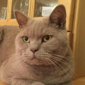
Young Winston is featured this week as he has fortunately made a swift and complete recovery following treatment for vomiting using an extended course of antacids and a bland diet. Apart from showing off his good looks this article illustrates a common feline problem and the approach taken for pets whose response to treatment is not quite so immediate as his.
You’ve probably witnessed your cat vomiting from time to time without raising too much concern. Vomiting is a protective mechanism that can result from something minor such as overindulgence or access to an irritant, or it can be a sign of a much more serious condition associated with a primary gastrointestinal disorder (e.g. an ingested foreign body) or a systemic disorder (e.g. kidney disease).
What is the difference between your cat vomiting and and your cat regurgitating? Why does it matter?
The oesophagus is a narrow, muscular tube that allows food to pass through on its way to the stomach. In healthy cats, food will move quickly through the oesophagus to the stomach with little to no delay. When you take your cat to your veterinary surgeon because he or she is vomiting, they will ask you questions in attempt to differentiate between vomiting and regurgitation.
Regurgitation is the passive ejection of contents from the oesophagus. The cat will lower its head and food is expelled with little or no effort. The food is usually undigested, may have a tubular shape, and is often covered with a slimy mucus. Cats will also attempt to eat the regurgitated material.
If the muscle of the oesophagus is considered diseased, it will either result in widening of the oesophagus due to loss of muscular tone (called ‘megaoesophagus’), or narrowing of the oesophagus, which acts as an obstruction to material moving down into the stomach (e.g. a stricture, a tumour or a foreign body can all cause narrowing of the oesophagus). A dilated (widened) oesophagus will not effectively, or efficiently, move or push food from the oesophagus into the stomach. This delay can result in regurgitation shortly after eating. The danger of regurgitation is that the contents may also be inhaled into the airways causing pneumonia and a cough. It’s therefore important for your vet to differentiate between vomiting and regurgitation as this will have an impact on deciding which diagnostic tests to perform, and also will help when deciding on the most appropriate treatment options.
Vomiting, on the other hand, is an active process. Cats will often vocalise, be apprehensive and heave/retch to vomit. If food is present in vomit, it is partially digested and can also contain a yellow fluid (bile). Vomiting can be divided down into primary (gastrointestinal) causes, or secondary (non-gastrointestinal) causes.
Primary causes of vomiting are those diseases directly affecting the stomach and upper intestinal tract. Secondary causes are due to diseases lying outside of the gastrointestinal tract and can include neurological disease or accumulation of toxic substances in the blood. Neurological disease and/or toxic substances will stimulate the vomiting centre in the brain and cause the animal to vomit. Additionally, vomiting can be further divided into acute versus chronic causes.
Some common causes for sudden (acute) vomiting in cats include:
- Diet-related causes (diet change, food intolerance)
- Gastric or intestinal foreign bodies (eg. toys, hairballs, or where one part of the intestine moves inside another part of the intestine)
- Gastrointestinal parasites
- Urinary tract causes: acute kidney failure, ruptured bladder
- Acute liver failure
- Pancreatitis
- Ingestion of toxins or chemicals
- Viral infections
- Certain prescribed medications
- Inner ear/neurological disorders
- Decompensation of a more chronic disease
Some common causes for chronic vomiting in cats (vomiting greater than 3 weeks duration) include:
- Gastritis/gastroenteritis (infectious, toxic, dietary indiscretion/intolerance)
- Hyperthyroidism
- Inflammatory bowel disease (gastritis, enteritis, colitis)
- Severe constipation
- Diabetes (ketoacidosis – this is a form of uncontrolled diabetes)
- Chronic liver disease
- Chronic kidney disease
- Pancreatitis
- Cancer (gastric/intestinal)
- Inner ear diseases/neurological disorders
- Heart worm disease
What should I do if my cat vomits frequently?
An occasional, isolated bout of vomiting is normal. However, frequent vomiting can be a sign of a more serious condition. Your vet will normally ask you a very detailed clinical history and perform a thorough physical examination. This information can help your vet narrow down potential causes from the very long list of possibilities.
The presence of fever, abdominal pain, jaundice, anaemia or abnormal masses in the abdomen will help your vet make a more specific diagnosis. The mouth should be carefully examined as some foreign objects such as string can entangle themselves around the base of the tongue with the rest of the object extending into the stomach or small intestine. A nodule may be palpated in the neck of cats with hyperthyroidism.
Many times, a full physical examination may be normal and unremarkable. At this stage, your vet may choose to adopt trial treatment/supportive care by implementing a brief starvation period, with or without administration of fluid therapy and various medications (e.g. pain relief, anti-nausea medications, antacids) and assess your cat’s response.
Further investigation of vomiting in cats
Depending on response, your vet may need to perform further investigations to differentiate primary from secondary causes of vomiting. Depending on history, clinical examination, and response to trial therapy, further tests may be needed and may include blood tests, urine analysis and faecal examination to rule out possible toxicities, parasites, and metabolic diseases. Further blood tests may include FIV/FeLV (Feline Immunodeficiency Virus and Feline Leukaemia Virus) to assess viral status. Although FIV/FeLV are not common primary causes of vomiting, they may reflect underlying immunosuppression which may make the cat more susceptible to certain diseases. Your vet may also decide to perform tests to assess the pancreatic health (e.g. pancreatitis).
Depending on the case, your vet may then decide to proceed with ‘second tier’ testing. This usually would involve some diagnostic imaging – x-rays and ultrasound – which can be useful in identifying masses, foreign objects, and other gastrointestinal tract problems such as pancreatitis.
Finally, if indicated, further ‘third tier’ testing may be indicated to obtain a biopsy of intestinal tract tissue if cancer or inflammatory bowel disease is suspected. Biopsies can be collected using either minimally invasive procedures (endoscopy and/or laparoscopy) or more invasive procedures (exploratory laparotomy). Endoscopy (a small camera attached to a long flexible tube) is commonly used to visualise the inside of the oesophagus, stomach and first part of the small intestine and to obtain small biopsies called ‘pinch biopsies’. It may also be possible to provide therapeutic interventions with an endoscope. Laparotomy (open surgery) or laparoscopy (‘key hole surgery’) may be considered for those cases where samples of organs lying outside of the stomach/small intestine and full thickness biopsies of intestines are needed.
This information will help your vet to identify the cause, streamline a treatment plan, and also provide you with a prognosis.
What are some treatment options?
The treatment for vomiting depends upon the underlying cause.
Non-specific treatment may include a 12-24 hour fasting period. Fluids may need to be withheld for a short period of time until vomiting has ceased. Water should never be withheld from an animal with known or suspected kidney disease without replacing fluids intravenously or subcutaneously (under the skin).
Water can be reintroduced in small volumes after a 6-12 hour period. You may wish to start with tuna water (not brine) or chicken broth (with NO added onion/garlic or powders) to encourage fluid intake. Gradual increases in volume may commence over the course of the day if vomiting does not recur.
Provided oral fluids have been well-tolerated, small volumes of a bland, high quality protein source (boiled chicken breast, tuna in spring water, turkey breast, boiled white fish) may then be introduced. It’s advisable to start with a teaspoon size portion every 4-6 hours for 1-2 days. Again, if tolerated, gradual increases in food volume may be attempted with the aim to wean back to the original diet over a 3-5 day period. If a cat is bright and alert and has had no previous health problems, episodes of acute vomiting may be managed at home, although veterinary consultation prior to home treatment is strongly advised. In certain situations (e.g. depression, dehydration, or symptoms lasting longer than 12-24 hours), your cat may require fluid therapy or drugs to help control vomiting. You’ll need to see your vet to determine the proper course of action.
What other symptoms should I watch for?
The causes of vomiting are so varied and can be extremely difficult to accurately diagnose. It’s therefore important to monitor for other signs of ill health that may help direct your vet in his/her quest in identifying the underlying cause.
What to watch for:
- Frequency of vomiting. If your cat vomits once and proceeds to eat regularly and have a normal bowel movement, the vomiting was most likely an isolated incident
- Diarrhoea
- Dehydration (sticky gums, weak, sunken eyes, increased ‘skin tenting’)
- Lethargy
- Blood in vomit
- Weight loss
- Change in appetite and water intake (either increased or decreased)
When is it time to see the vet?
Please see your vet if you notice any of the symptoms mentioned above or if vomiting persists. Depending on your cat’s age, medical history, physical examination findings and particular symptoms, your vet may choose to perform various tests and/or admit your cat for more intensive treatment.


