News
Archive for July, 2017
Special Offer – July 2017 – Dry Eye
by admin on July 4th, 2017
Category: Special Offers, Tags:
Pet of the Month – July 2017 – Henry
by admin on July 4th, 2017
Category: Pet of the Month, Tags:
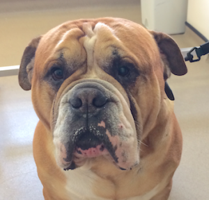
Our pet of the month is Henry who we are pleased to say has recovered fully from a severe bout of pancreatitis, which necessitated a period of hospitalisation.
What is pancreatitis?
The pancreas is an organ in the abdomen (tummy) which is responsible for releasing enzymes (types of proteins) to digest food. The pancreas also releases important hormones (such as insulin) into the bloodstream. Pancreatitis occurs when the pancreas becomes inflamed (tender and swollen). In most cases pancreatitis occurs for no apparent underlying reason, although sometimes it can have a particular cause (such as scavenging food). Pancreatitis most commonly affects middle aged to older dogs, but in addition, dogs of certain breeds (e.g. Cocker Spaniels and Terrier breeds) are more prone to developing the condition.
What are the signs of pancreatitis?
Pancreatitis can cause a variety of symptoms, ranging from relatively mild signs (e.g. a reduced appetite) to very severe illness (e.g. multiple organ failure). The most common symptoms of pancreatitis include lethargy, loss of appetite, vomiting, abdominal pain (highlighted by restlessness and discomfort) and diarrhoea.
How is pancreatitis diagnosed?
The possibility that a dog may be suffering from pancreatitis is generally suspected on the basis of the history (e.g. loss of appetite, vomiting) and the finding of abdominal pain on examination by the veterinary surgeon. Because many other diseases can cause these symptoms, both blood tests and an ultrasound scan of the abdomen are necessary to rule out other conditions and to reach a diagnosis of pancreatitis. Although routine blood tests can lead to a suspicion of pancreatitis, a specific blood test (called ‘canine pancreatic lipase’) needs to be performed to more fully support the diagnosis. An ultrasound scan is very important in making a diagnosis of pancreatitis. In addition, an ultrasound scan can also reveal some potential complications associated with pancreatitis (e.g. blockage of the bile duct from the liver as it runs through the pancreas).
How is pancreatitis treated?
There is no specific cure for pancreatitis, but fortunately, most dogs recover with appropriate supportive treatment. Supportive measures include giving an intravenous drip (to provide the body with necessary fluid and salts) and the use of medications which combat nausea and pain. Most dogs with pancreatitis need to be hospitalised to provide treatment and to undertake necessary monitoring, but patients can sometimes be managed with medication at home if the signs are not particularly severe. At the other extreme, dogs that are very severely affected by pancreatitis need to be given intensive care.
One of the most important aspects of treating pancreatitis is to ensure that the patient receives sufficient appropriate nutrition while the condition is brought under control. This can be very difficult because pancreatitis causes a loss of appetite. In this situation, it may be necessary to place a feeding tube which is passed into the stomach, and through which nutrition can be provided (see Information Sheet on Feeding Tubes). If a dog with pancreatitis is not eating and will not tolerate a feeding tube (e.g. due to vomiting), intravenous feeding (using a drip to supply specially formulated nutrients straight into the bloodstream) may be necessary.
What is the outcome in pancreatitis?
It may be necessary for dogs with pancreatitis to be hospitalised for several days, but fortunately, most patients with the condition go on to make a complete recovery, provided that appropriate veterinary and nursing care is provided. In some instances, dogs can suffer repeated bouts of the condition (called ‘chronic pancreatitis’) and this may require long term management with dietary manipulation at the forefront.
Disc Disease in Dogs
by admin on July 4th, 2017
Category: News, Tags:
Dachshunds and similar breeds such Pekingese, Lhasa Apsos and Shih Tzus, have short legs and a relatively long back. These breeds suffer from a condition called ‘chondrodystropic dwarfism’. As a result they can be prone to back problems, more specifically disc disease.
What are intervertebral discs?
The intervertebral disc is a structure that sits between the bones in the back (the vertebra) and acts as a shock absorber. The structure of a disc is a little like a jam doughnut with a tough fibrous outer layer (annulus fibrosus) like the dough, and a liquid/jelly inner layer (nucleus pulposus) like the jam.
In chondrodystrophoid breeds like dachshunds, the discs undergo change where the nucleus pulposus changes from a jelly-like substance into cartilage. Sometimes the cartilage can mineralise and become more like bone and show up on an x-ray. In these breeds, this change occurs in all discs from approximately one year of age and is considered part of usual development for these dogs. This change in the discs is a degenerative process and predisposes the disc to disease.
The most common type of disc disease in dachshunds and other chondrodystrophoid breeds is ‘extrusion’. This is where the annulus fibrosus ruptures and allows the leakage of the nucleus pulposus into the spinal canal. An analogy we often use is the jam leaking from a jam doughnut when it is squeezed.
What are the signs of disc disease in dachshunds and other breeds?
The first evidence that there might be a problem is that your dog may be in pain. This may consist of yelping and/or a hunched back and more subtle signs such as quietness and inappetence (lack of appetite). Back pain is commonly mistaken for abdominal pain.
After pain there may be mobility problems. The first thing to occur is wobbliness of the hind limbs – termed ‘ataxia’. After this, weakness can develop with the wobbliness, which is termed paresis. Subsequently, the patient may lose control of the back legs altogether and not be able to walk. If there is no movement in the hind limbs this is ‘paralysis’.
Will my dog need to be referred to a specialist?
Spinal disease can sometimes be difficult to manage and your dog may be referred to a specialist. At the referral clinic, your dog will be assessed by the specialist neurologist or neurosurgeon. They will test various reflexes to decide where they think the issue may be. They will perform a clinical examination which will assess for other disease processes and look for any areas of pain. One thing that is important to assess is the presence of pain sensation. We call this ‘deep pain sensation’. To assess this, the specialist will pinch your dog’s toes, usually with fingers initially but it may need to be with forceps. This is not a pleasant test to perform but it is necessary as the presence of deep pain comes with a more favourable prognosis. The loss of deep pain sensation means the prognosis is worse and without surgery is considered very poor.
What investigations will be performed?
After the clinical examination, we will generally have a good idea where the problem is and the next step is to confirm the diagnosis. The best way of diagnosing disc disease is with MRI (magnetic resonance imaging) or CT (computed tomography). With MRI we get detailed information about the soft tissue such as the disc itself and the spinal cord. Computed tomography uses the same technology as x-ray and provides good information about the bone and mineral structures and can tell us where the lesion is and whether or not surgery is indicated.
How is disc extrusion treated?
If a disc extrusion – the ‘jam out of the doughnut’ scenario – is diagnosed then we can opt for conservative care or surgical management.
Conservative care is a non-surgical management and can be considered in dogs where the signs are mild or intermittent and when there is only mild compression of the spinal cord. It can also be considered if there are concerns regarding the cost of surgery.
Surgical treatment involves making a window through the bone structures of the vertebra, into the spinal canal, a procedure called a ‘hemilaminectomy’. Once into the spinal canal, the disc material (nucleus pulposus) is removed and the spinal cord is decompressed. Surgical treatment tends to give a speedier and more predictable recovery and is often considered the treatment of choice, where indicated.
What is the prognosis for a dog with disc disease?
When dealing with spinal injury, particularly from disc disease, we aim for patients to achieve a functional recovery. This means the patient is pain free, is able to get around on their own (although they may still be wobbly) and able to toilet themselves unaided. Whether a patient will return to normal or whether there will be residual deficits is down to the individual.
The prognosis for patients who have intact deep pain sensation is favourable, with most patients (80-90%) achieving a functional recovery. In patients without deep pain sensation, the prognosis is guarded, with only 50% of cases achieving a functional recovery but without surgery the prognosis for a functional recovery is approximately 5%.
Surgery tends to speed recovery and aid early pain control. We expect most patients to recover within the first 2 weeks after surgery, with a smaller number of patients taking longer (2-6 weeks). If there is no improvement after 6 weeks the prognosis becomes more uncertain and long term disability may be present. This occurs in approximately 1 in 10 patients.


